Breakthrough in Blood Flow Imaging: PACTER
Good news in vascular health is on the horizon! Discover PACTER, the innovative 3D imaging breakthrough poised to elevate blood flow analysis to new heights of safety and precision in our latest uplifting article.
Introduction to Advanced Hemodynamics Imaging
A collaboration between Caltech and the University of Southern California has led to a groundbreaking development in medical technology, as reported by the National Institute of Biomedical Imaging and Bioengineering. Their new imaging method, known as Photoacoustic Computed Tomography through an Ergodic Relay (PACTER), is poised to transform how clinicians monitor and diagnose vascular diseases.
Why It Matters
Blood flow imaging is crucial for detecting strokes, peripheral artery disease, and diabetic vascular complications. Improving safety and precision could mean faster, more accurate diagnoses and better outcomes for patients.
The Need for Safer Imaging Techniques
Current imaging methods often require the use of potentially harmful contrast agents or exposure to ionizing radiation. These risks have prompted researchers to seek safer, non-invasive alternatives for visualizing the intricate details of blood flow, crucial for detecting and managing vascular conditions.
How PACTER Works
PACTER relies on the photoacoustic effect, a process where light is absorbed and re-emitted as sound waves. This innovative system uses just one sensor to create 3D images, circumventing the need for multiple expensive sensors and thereby making the technology more accessible.
Calibration: The Key to PACTER's Success
The research team's calibration strategy is at the heart of PACTER's effectiveness. By directing laser beams at thousands of points in a blood sample, they have enabled the system to:
Distinguish between 6,400 signals from a single laser beam during imaging
Decompose raw signals into individual components
Utilize a single sensor as if it were thousands of virtual detectors
Achieve rapid data collection for real-time imaging
Demonstrating PACTER's Capabilities
The team has successfully applied PACTER in animal studies and human trials. In mice, the system mapped abdominal blood vessels and breathing rates. In humans, it assessed blood flow in areas affected by peripheral vascular disease and diabetes, proving its potential for clinical use.
The Future of PACTER Technology
While the current iteration of PACTER marks a significant achievement, ongoing improvements are anticipated. Senior author Lihong Wang notes the system's cost-effectiveness and the team's aspirations for enhancing sensitivity and portability. Prospective applications include:
Measuring blood oxygenation in neck arteries and veins
Tracking metabolism in the brain
Continuous monitoring for hospital and home healthcare
Implications for Clinical Practice and Research
This technology's advancement underscores the importance of basic research in leading to clinical breakthroughs. The researchers hope that the study will inspire further exploration into PACTER's vast potential and lead to new solutions for vascular health challenges.
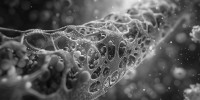
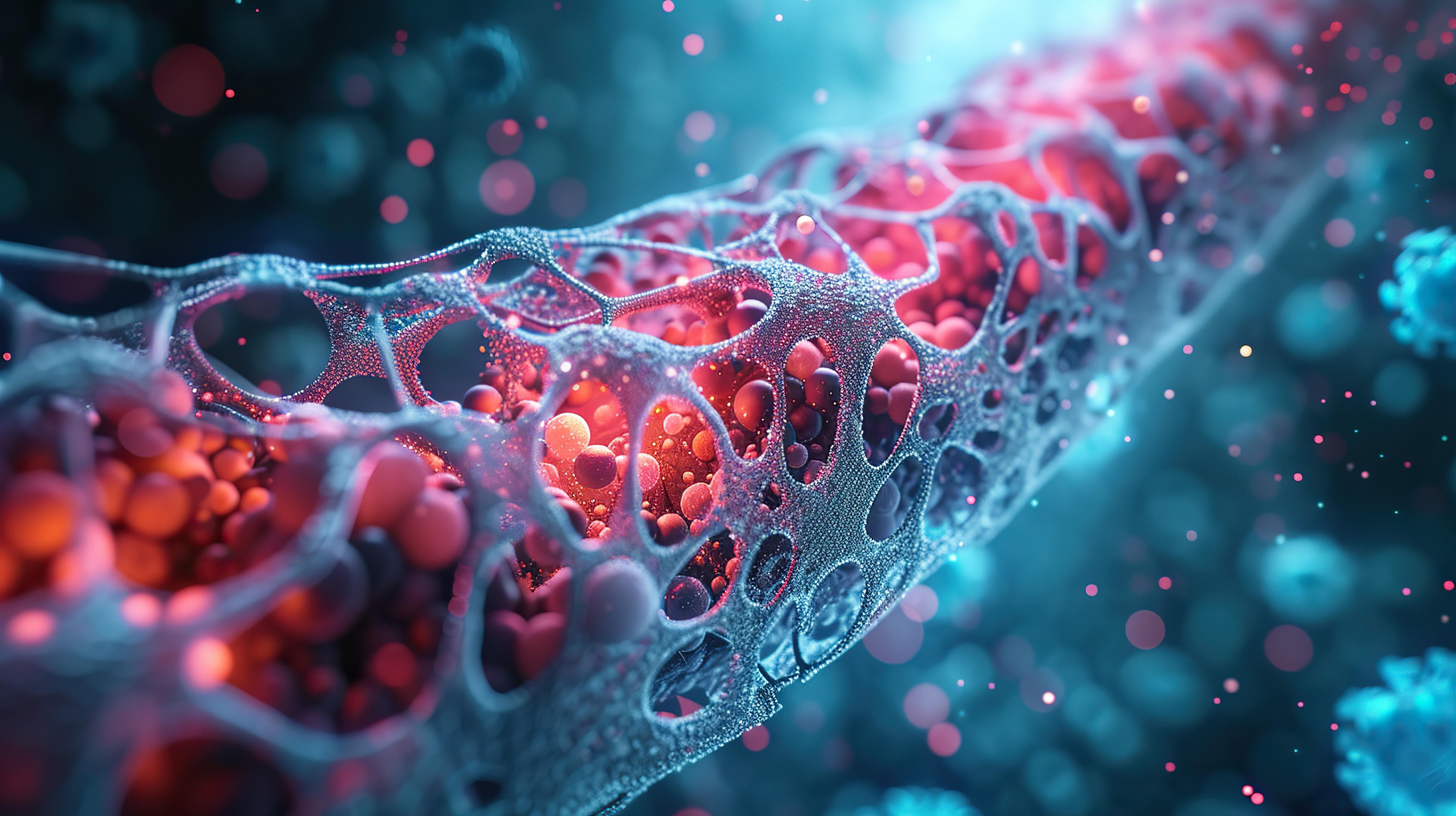
 Single-shot volumetric photoacoustic imaging using a single-element detector may facilitate the early detection and monitoring of peripheral vascular diseases and may be advantageous for use in biometrics and point-of-care testing.
Get to the study: Funded by grants from the NIBIB and the National Cancer Institute, the study represents a milestone in medical imaging research. For those interested in the detailed findings, the study by Yide Zhang et al. is available in Nature Biomedical Engineering (2023), here.